Nissen R
Hernia (textbook) 1964;30:488-96.
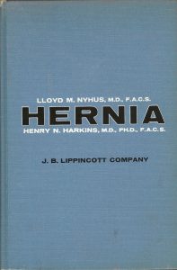
Nissen, R. (1964). The treatment of hiatal hernia and esophageal reflux by fundoplication (chapter 30). In L.M. Nyhus & H.N. Harkins (Eds), Hernia (1st ed., pp.488-96). Philadelphia, PA: Lippincott.
After searches in PubMed.gov, Amazon.com and AbeBooks.com we were unable to locate a digital copy of this book chapter. The following material may be protected by copyright law.
It is our intent to honor Rudolf Nissen, MD’s work by highlighting the text from the book chapter’s introduction. In it, Dr. Nissen describes the procedure that bears his name: Nissen fundoplication. Dr. Nissen’s approach is fundamental to how hiatal hernia and gastroesophageal reflux disease (GERD) is treated. In PubMed.gov, Nissen R is associated with 212 publications so his work is referenced heavily for this disease state and surgical techniques.
Section: Diaphragmatic Hernia, Esophageal Hiatus
Chapter 30: The Treatment of Hiatal Hernia and Esophageal Reflux by Fundoplication
Rudolf Nissen
“A stitch in time saves nine.” ~Anon.
Introduction
The clinical manifestations of the hiatal hernias which require surgical treatment, were due in 80 per cent of our cases to a functional insufficiency of the gastroesophageal sphincter and its sequelae. Consequently, the principal purpose of the operation is not the elimination of the hernia, but the restoration of sphincteral function. Presumably this principle is now generally accepted.
As regards the causal relationship between reflux esophagitis and a sliding hiatal hernia, there is still some controversy, since reflux symptoms without a hernia and hernias without reflux symptoms are occasionally observed. In the former instances there almost always exists a so-called cardiofundal defect (a funnel-shaped transformation of the cardia, with the formation of a wider gastroesophageal angle), a malformation which we regard as the preliminary stage of a true sliding hernia. And just as may the latter condition, in the more severe cases it too many lead to reflux esophagitis. Hernias without reflux are usually asymptomatic. If symptoms are present, they frequently are incorrectly attributed to the hernia.
The most unequivocal roentgenologic and clinical manifestations of regurgitation are observed after a gastroespohageal resection.
In 1936 we performed a cardial resection in a 28-year-old male with an ulcer in the distal portion of the esophagus, which had penetrated into the pericardium. Since we then were unaware of the published work of Sea and Ohsawa, we believed that it was the first instance of a successful one-stage transthoracic cardial resection, and therefore published the case. Since in previous unsuccessful attempts failure was due to the inadequacy of the sutures in the gastroespohageal anastomosis, we attributed the inadequate healing of the surgical wound to the anastomosis technic (a simple apposition of thee stumps). It later was found that this was not the only factor and certainly not the most important one. At that time we attempted to prevent such an inadequacy by embedding the esophageal stump in the wall of the remaining portion of the stomach, as the rubber tube is burned in the Witzel type of gastrostomy. Control studies which we were able to make in this patient 16 years later showed that not only had there been no inadequacy in the healing of the anastomosis, but that there had been no occurrence of the reflux esophagitis almost always previously encountered in such cases.
When an evaluation of our results of the conventional surgical methods of treating hiatal hernia revealed that approximately 50 per cent were clinically and/or roentgenologically failures, we tried making the elimination of the reflux the chief goal of corrective surgery. This was done at first by means of gastropexy, a procedure which had been under test since 1947 and was first described in the literature in 1954. It consists of a small transabdominal operation in which the lesser curvature of the stomach is sutured under tension to the anterior abdominal wall. This results in an eversion of the hernial sac and the establishment of a very acute angle at the junction of the esophagus with the stomach. The long-term results of gastropexy in our hands were definitely better than those achieved with the various types of hernioplasty. In 81 per cent of our patients treated by this method the reflux symptoms disappeared permanently. Boerema, who began to perform gastropexies independently of us in 1955, had approximately the same results. However, after 5 years’ observation time there was a gradually increasing rate of roentgenologically demonstrable recurrence, which reached 35 per cent (half of them asymptomatic).
In the meantime a series of patients with therapy-resistant reflux esophagitis without roentgenologically recognizable hiatal hernia was assigned to us. Encouraged by the above-described good long-term findings in the patient who had had a gastroesophageal resection, we believed that we would be able to solve the problem of the reflux of gastric juice in a simple manner if in imitation of the embedding of the esophageal stump in the gastric wall we mobilized the distal portion of the esophagus transabdominally, wrapped the fundus around it, and fixed it in this position with a serious of sutures. This operation, which has been termed fundus-enfolding or fundoplication, was performed for the first time in 1955, and after confirmation of its success in a small series of patients, a description of it was published in 1956. We were so impressed by the prompt and lasting results of this relatively simple operation that we then began using both fundoplication and gastropexy in all hiatal hernias accompanied by reflux symptoms. When experience had shown that fundoplication as the sole surgical procedure caused disappearance of the symptoms of a sliding hernia with the same reliability, gastropexy was abandoned in favor of fundoplication in all hiatal hernias except those of the para-esophageal type. In the meantime, the operation had been performed in 260 cases.


